I am...

Langerhans Cell Histiocytosis in Children

Langerhans cell histiocytosis (LCH) is the most common of the histiocytic disorders and occurs when the body accumulates too many immature Langerhans cells, a subset of the larger family of cells known as histiocytes.

Langerhans cell histiocytosis (LCH) is the most common of the histiocytic disorders and occurs when the body accumulates too many immature Langerhans cells, a subset of the larger family of cells known as histiocytes. Histiocytes are cells within the immune system that function as “phagocytes,” which means they ingest and remove foreign bodies like bacteria and debris. They were first discovered by Paul Langerhans in 1868. These cells are normally found in the skin, bone marrow, blood, liver, lungs, lymph glands and the spleen. Langerhans cells are a type of white blood cell (immune cells) that normally help the body fight infections.  In LCH, too many abnormal Langerhans cells are produced, together with other types of inflammatory cells, and these cells build up in certain parts of the body where they can form tumors or damage organs.

In 2021, the National Comprehensive Cancer Network published clinical guidelines for histiocytosis classifying LCH, ECD (Erdheim-Chester disease), and RDD (Rosai Dorfman disease) as histiocytic neoplasms. LCH is considered a form of cancer with lots of inflammation-like symptoms (or inflammatory neoplasia). It is classified as a blood cancer by the World Health Organization (WHO). Indeed, some forms of LCH can be treated with chemotherapy (cancer therapies) and are treated by doctors who treat cancer patients (oncologists). However, this doesn’t necessarily mean that all of the worrisome implications that come along with the term “cancer” are true of LCH for every patient; LCH and other histiocytic disorders have a wide spectrum of presentations.

The Histiocytosis Association works closely with the Histiocyte Society, an international organization of over 240 physicians and researchers, which is dedicated to studying the histiocytic disorders. Through this partnership, understanding of the disease has greatly increased, and survival rates and quality of life continue to improve. The information on this site is reviewed and updated regularly by members of the Histiocyte Society with oversight by the Histiocytosis Association’s Board of Trustees’ Scientific Commitee.

The information on this page has been written and reviewed by the Histiocytosis Association Board of Trustees Scientific Committee and a member of the Histiocyte Society, and subsequently audited by patients and families to ensure enough information was captured. The most recent update to this page was in January of 2025.

Cause of LCH

LCH is caused by somatic mutations (gene changes acquired after birth and only present in certain cells) in genes that control how histiocytes, or LCH cells, function. These include mutations of the BRAF, MAP2K1, RAS, and ARAF genes. These mutations may cause too many LCH cells to grow in areas where they are not usually present, and damage tissues or form lesions (tumors).

The BRAF gene provides instructions for making a protein that controls cell growth and development. The BRAF protein can be switched on and off in response to some chemical signals. Somatic mutations will cause the BRAF protein in involved cells to be continuously turned on, and to send messages to the nucleus even without any chemical signals. This overactive protein will then lead to the development of LCH by causing the Langerhans cells to divide without control and grow. Because somatic gene mutations are not present in the germ cells (egg and sperm), they are not passed on from parents to their children (are not inherited).

The BRAF gene, mutated in about half of people with LCH, is part of a family of genes called oncogenes. When changed (mutated), oncogenes can allow normal cells to become cancerous. This is why today, LCH is being considered as a form of cancer with lots of inflammation-like symptoms (or inflammatory neoplasia). It is classified as a blood cancer by the World Health Organization (WHO). Indeed, some forms of LCH can be treated with chemotherapy (cancer therapies) and are treated by doctors who treat cancer patients (oncologists). Although some forms of LCH may behave like cancer, most forms will have a very benign course and many of these can be observed without any treatment. In other words, most patients with LCH will survive this disease.

History of LCH

Histiocytosis was first described in the medical literature in the mid to late 1800s. Through the years, it has been known by various names, such as, eosinophilic granuloma, Letterer-Siwe disease, Hashimoto-Pritzker disease, and Hand-Schüller-Christian syndrome and histiocytosis X . While these terms all refer to LCH, they described specific patterns of LCH that, at one time, seemed to be different diseases. In 1987, the name Langerhans cell histiocytosis (LCH) was introduced as a term that described all of those names with just one diagnosis. This name was agreed upon to recognize the central role of the Langerhans cell.

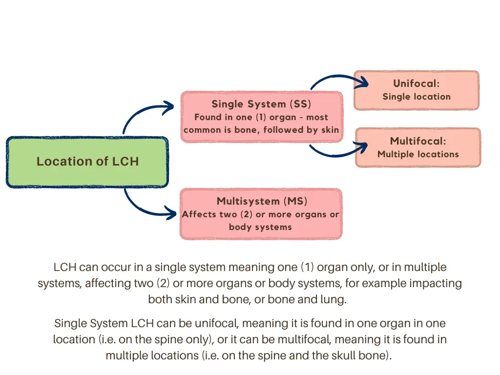
LCH is believed to occur in 5 per million children per year, but any age group can be affected, from infancy through adulthood. In newborns and very young infants, it occurs in 1-2 per million each year. It is, however, believed to be under-diagnosed, since some patients may have no symptoms, while others have symptoms that are mistaken for injury or other conditions. It occurs most often between the ages of 1-3 years and may appear as a single tumor, or “lesion,” (area of abnormal tissue or tumor) or can affect many body systems, such as skin, bone, lymph glands, liver, lung, spleen, brain, pituitary gland and bone marrow.

Information has been collected in various studies which show that bone involvement occurs in approximately 78% of patients with LCH and often includes the skull (49%), hip/pelvic bone (23%), upper leg bone (17%) and ribs (8%). Skin LCH is seen in as many as 50% of patients. LCH within the lung is seen in 20% to 40% of patients, while 30% of patients have lymph node involvement.

Symptoms depend on the location and severity of involvement. LCH is usually diagnosed with a tissue biopsy, (when a sample of tumor material is taken with a medical or surgical procedure), in addition to other testing, such as x-rays and blood studies. A biopsy of a suspected LCH tumor is necessary to make a definitive diagnosis.

Some cases of LCH causing little or no symptoms or health problems may not require treatment; for patients with more extensive disease, chemotherapy may be necessary. Hematologists and oncologists, who treat cancer, are most frequently the health care providers who treat children with Langerhans cell histiocytosis.

LCH in the skin, bones, or lymph nodes is called “low-risk” because it most often gets better without treatment. Some patients have LCH in the spleen, liver, and bone marrow; this is called “high-risk-disease” and may be more difficult to treat. LCH affecting the brain is rare but can be very serious. Some LCH patients may develop long-term side effects such as diabetes insipidus, stunted growth, loss of teeth, bone defects, hearing loss, or neurologic problems; while other patients remain without side effects (more details about this are below). In a minority of cases, the disease can be life-threatening.

Patients with LCH should usually have long term follow-up care to detect late complications of the disease or treatment. It is recommended to speak with your treating physician about a long-term follow up plan and options for your child as they transition to adulthood to maintain contact with a histiocytosis physician.

Classification of LCH

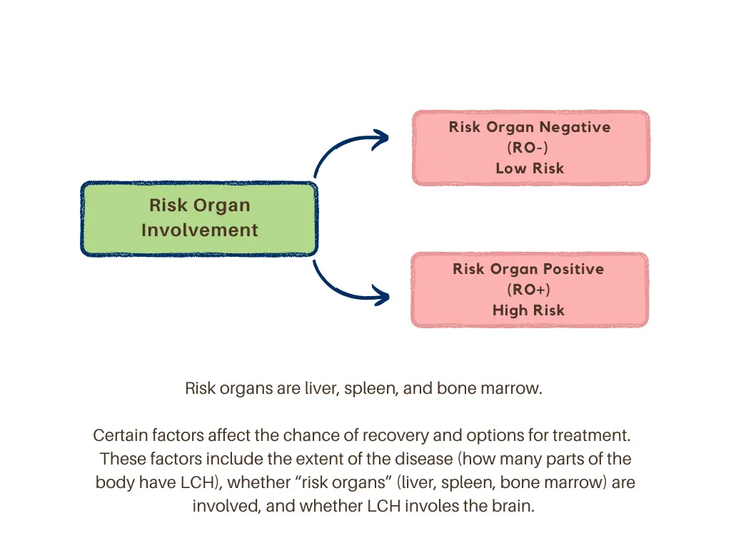
Certain factors affect the chance of recovery and options for treatment. These factors include the extent of the disease (how many parts of the body have LCH), whether “risk organs” (liver, spleen, bone marrow) are involved, and whether LCH involves the brain. Also, whether LCH is improving within the first few weeks of treatment gives some indication about the future outlook.

Patients with LCH need ongoing, long-term follow-up and surveillance with health care providers to watch for late complications of either LCH itself, or of LCH treatment. These may include problems with bone deformity, or bone function, liver or lung problems, endocrine (hormone) abnormalities, dental issues, neurological (speech and balance), chronic pain or fatigue, or cognitive (learning, memory) problems. You should discuss a plan for long-term follow up with your current treating physician, and seek guidance about involving other physicians with specialized expertise into your long-term care team.

Symptoms

Langerhans cell histiocytosis (LCH) may be limited to only one part of the body, such as a bone, skin, or it can involve many organs.The disease may be more serious in young infants or if “risk” organs such as liver, spleen, or bone marrow are involved.

The following symptoms may indicate disease involvement of a particular organ, however, only with a biopsy of the impacted area(s) can a diagnosis of LCH be confirmed. This disease varies greatly from patient to patient, and some but not all of these symptoms may be present:

  • Bone (single or multiple sites)
    • Swelling or a lump, with or without pain, of a bone like the skull, bones around eyes or ears, jaw bone, spine, arms or legs, ribs or hips
    • Headaches, fractures, limp, trouble walking, neck or back pain (collapsed vertebra or vertebra plana)
    • Children with LCH lesions in bones around eyes, ears, or jaws have a higher risk of developing late Diabetes Insipidus and/or other central nervous system complications
  • Skin
    • Can be the only site affected in infants. In few cases, skin only LCH may get worse and evolve into a form called high-risk multisystem LCH
    • Scalp rash that can manifest like a “cradle cap” in infants or dandruff in children or adults
    • Groin rash, armpits rash, oozing, tenderness, redness, or itchy painful rash around the arms; rash may also occur in the abdomen, back, or chest
    • Decoloration and hardening of nails, loss of nails
  • Mouth
    • Swollen gums, or teeth that fall out or are uneven
    • Sores on the tongue or lips, or inside the cheeks
  • Bone Marrow
    • Low red blood cells (anemia) with pale skin, feeling tired, and decreased appetite
    • Low white blood cells; frequent infections, fevers
    • Low platelets (clotting cells); easy bruising or bleeding
  • Gastrointestinal Tract (stomach/intestines/colon)
    • Abdominal pain, vomiting, diarrhea, blood in stool
    • Child’s growth falls below the standard expectation because of low nutrition
  • Liver/Spleen
    • Swelling of abdomen caused by a big liver and/or spleen or buildup of extra fluid
    • Yellow skin and eyes; itching, feeling very tired
    • Easy bruising/bleeding, bloody stools, diarrhea
  • Endocrine System (hormones)
    • Pituitary gland: strong thirst and frequent urination (diabetes insipidus), slow growth, early or late puberty, weight gain
    • Thyroid gland: swollen gland, hypothyroidism that can cause lack of energy, constipation, dry skin, thinning of the hair, being sensitive to cold, depression, trouble concentrating; loss of appetite or choking on food in infants; behavior problems, weight gain, slow growth and late puberty in children and adolescents
  • Lung
    • Chest pain, dry cough
    • Trouble breathing (especially in adults who smoke)
    • Coughing up blood
    • Collapsed lung: chest tightness, feeling tired, bluish color to the skin
  • Central Nervous System (CNS): brain or spinal cord
    • Trouble walking, loss of balance, uncoordinated body movements
    • Trouble speaking or seeing
    • Headaches, dizziness, seizures
    • Changes in behavior, memory and learning problems
    • All of the above symptoms can be caused by lesions in the central nervous system or by central nervous system neurodegenerative syndrome
  • Ears
    • Discharge from ear canal, redness, itchy rash at the external ear canal
    • Ear pain that can mimick recurrent ear infections
    • Reduction or (rare) loss of hearing
  • Eyes
    • Bulging of eyeballs, swelling above the eyes
    • Vision problems and (rare) blindness
  • Lymph Nodes
    • Neck, groin, armpits: swollen nodes, tenderness
    • If the enlargement surrounds the respiratory area (mediastinum), it can result in cough, shortness of breath, or turning blue

The kidneys and gonads (ovaries and testicles) are usually not affected by LCH.

The organs and tissues most commonly involved are the bones (including eye bones), skin, lymph nodes, bone marrow, lungs, pituitary gland, spleen, liver, and digestive tract.Involvement of more than one system is common.For example, bones can be affected alone or as part of multisystem disease.Lymph nodes and skin involvement also can be either single-system disease or part of multisystem LCH.Young infants with skin involvement should be followed closely by a knowledgeable physician (a pediatric oncologist), as some cases can spread to vital organs and quickly become life-threatening.Central nervous system (or brain) involvement (CNS), including the pituitary gland, is often part of multisystem (extensive) disease, although there are cases in which diabetes insipidus is the first presenting symptom.

It is important to remember that symptoms alone do not diagnose LCH, and a biopsy of an involved site is necessary.Thus, it is important to consult a physician to receive an appropriate workup and accurate diagnosis.

Diagnosis & Treatment

There have been several pediatric clinical trials for LCH which have given a wealth of understanding on treating children. Information about diagnosis and treatment below is updated regularly to include all of these options and new options as well.

The National Comprehensive Cancer Network® (NCCN®) – an alliance of leading cancer centers – announced the publication of new NCCN Guidelines® for histiocytosis. These clinical practice guidelines provide the latest evidence and expert-consensus for diagnosing and treating the three most common forms of histiocytosis in adults: Langerhans cell histiocytosis (LCH), Erdheim-Chester Disease (ECD,) and Rosai Dorfman Disease (RDD). Although the guidelines are focused on adult patients, there may be insight for pediatric physicians as well. The Guidelines are listed as “histiocytic neoplasms” and can be found on NCCN’s website.

You can find international expert consensus recommendations for the diagnosis and treatment of Langerhans cell histiocytosis in adults, here. You can download a copy, here. It is encouraged that you share this with your physicians for reference.

On November 1st, 2022, Memorial Sloan Kettering Cancer Center (MSK) announced that the U.S. Food and Drug Administration (FDA) has approved the oral MEK inhibitor drug cobimetinib (Cotellic®) for the treatment of adult patients with the family of blood diseases known as histiocytic neoplasms (HN). These diseases include Erdheim-Chester disease, Rosai-Dorfman disease, and Langerhans cell histiocytosis. Cobimetinib is an oral inhibitor of MEK1 and MEK2, currently approved to treat melanoma. Gratitude and congratulations to the Principal Investigator of this study, Dr. Eli Diamond, neuro-oncologist and neurologist at MSK and Chair of the Scientific Committee for the Histiocytosis Association. We applaud the efforts of Dr. Diamond and the entire study team for this tremendous advancement. Read more here.


Diagnosis

The diagnosis of LCH is made following a biopsy of the affected tissue. A small piece of the tissue is obtained so that it can be viewed under a microscope by a pathologist. If the cells in the tissue have expression of specific proteins (Langerin, CD1a), the diagnosis of LCH can be made. This procedure can be performed on any location but usually is done on the skin, bone or lymph nodes.

In addition, the following tests may be performed depending on particular symptoms in the child.

  • Physical examination and history: Recording of height, weight, temperature, pulse, family history, history of symptoms, past illnesses, etc.
  • Neurological examination: Testing of ability to walk, muscle testing, coordination, and cognition (attention, memory, learning ability).
  • Complete blood count (CBC): Checking levels of red blood cells, white blood cells, and clotting cells (platelets).
  • Blood chemistry tests: Studies for kidney, coagulation or clotting, thyroid and immune function; abnormal level can be a sign of disease.
  • Liver function tests: Checking blood levels of substances released by the liver; an abnormal level can be a sign of disease in the liver.
  • Urinalysis: Testing the amount of red cells, white cells, protein and sugar in the urine.
  • Water deprivation test: Checks how much urine is made and whether it becomes concentrated when water is withheld; used when diabetes insipidus is suspected.
  • Biopsy: The removal of tissues so that they can be viewed under a microscope by a pathologist to check for LCH cells. To diagnose LCH, a biopsy of bone, skin, lymph nodes, liver, or other sites of disease may be performed.
  • Bone marrow aspirate and biopsy: The removal of bone marrow, blood and a small piece of bone by inserting a hollow needle into the hipbone; the sample is studied under a microscope to look for signs of LCH.
  • BRAF and MAPK gene testing: A lab test in which a sample of tissue or blood is tested for changes in the BRAF or other MAPK pathway genes. (Molecular testing)
  • X-rays: Performed of the lungs (chest) or of all the bones of the body (skeletal survey) to determine if there are abnormalities.
  • Bone scan: A scan for bone lesion in which a small amount of radioactive material is injected into a vein, collects in any abnormal parts of the bones and is shown on a scanner. This type of scan is rarely performed anymore in practice.
  • CT scan: For this x-ray dye may be injected into a vein or swallowed to help find any abnormalities; the scan takes a number of detailed pictures at different angles inside the body.
  • PET scan: A small amount of radioactive sugar is injected into a vein, and diseased cells show up brighter on the scanner. This scan is usually combined with a CT scan.
  • MRI: Using a substance called gadolinium injected into a vein to highlight areas of disease involvement, this scan then takes a number of detailed pictures inside the body.
  • Ultrasound: High-energy sound waves are bounced off organs and tissue and make echoes, which form a picture of the internal body.
  • Endoscopy: When involvement of the digestive tract is suspected, a tube is passed into a body opening such as mouth or rectum to look at internal tissue; often, biopsies are also taken at the same time.

Treatment

Treatment of LCH depends upon the individual patient and is planned after thorough testing to determine the extent of disease.

  • Single System (SS) LCH:
    • In some cases with limited or single organ involvement (such as single bone or skin only), LCH may regress (improve or resolve) on its own without treatment. In others, minimal treatment such as surgical removal (or curettage—scraping–of a single bone lesion), anti-inflammatory drugs (indomethacin) for single or multiple bone lesions, or steroids (creams or pills) for skin disease, will result in a favorable outcome.
    • Prednisone is a steroid medication which inhibits the function of white blood cells, like lymphocytes, and macrophages. Since these inflammatory cells accompany the histiocytes in LCH lesions, the treatment effect in LCH could be directly on the LCH tumor cells or the surrounding cells. Oral medications like methotrexate, 6-mercaptopurine (6-MP) and thalidomide have been used to treat isolated skin disease.
  • Multisystem (MS) LCH:
    • In patients with more extensive disease (multisystem), systemic chemotherapy is most often necessary. Several drugs and combinations have been shown in clinical trials to give excellent outcomes with minimal side effects.
    • The most commonly studied chemotherapy drugs in LCH are oral prednisone and intravenous vinblastine combinations. Vinblastine is the salt of an alkaloid extracted from a plant that works by inhibiting the movement of chromosomes between dividing cells thus causing the LCH (or tumor cells) to break and die. It is generally well tolerated and has less neurotoxicity than vincristine. This means the patient will have less intense foot/hand cramps or tingling, less trouble walking and less constipation.
    • For patients with severe extensive disease that comes back after initial response, or not responsive to initial treatment with prednisone/vinblastine, chemotherapy with intravenous cladribine (2-CdA), or clofarabine, or cytarabine, have shown promising results. These drugs work by similar mechanisms of substituting the drug for a building block of DNA synthesis, thus causing the LCH cells to die.
      • Cytarabine and cladribine are equally effective for low-risk patients (90% responses and few relapses), while cladribine alone was not as effective in high-risk patients (40% response rate).
      • Clofarabine has usually been reserved for patients whose LCH came back after the treatment was done (relapse or reactivation) with cladribine or cytarabine; long-term responses occur in 80% of patients. However, the drug is very expensive and many insurance companies may not approve it unless the patient has failed few prior treatments.
  • Hydroxyurea (Hydrea):
    • Hydroxyurea inhibits construction of the DNA strand backbone and has been used to treat diseases of myeloid cells in the bone marrow (chronic myeloid leukemia or CML) for decades. Since LCH is caused by a BRAF-V600E (or other gene) mutations of the myeloid cells of the bone marrow, hydroxyurea was found to be a reasonable treatment choice. It was found to be effective for LCH involving the skin, oral cavity, external ear canal or single bone lesions.
  • BRAF Gene Inhibitors:
    • LCH patients with the BRAF gene mutation respond very well to BRAF inhibitors (dabrafenib, vemurafenib), and/or MEK inhibitors (trametinib, cobimetinib), especially those with high-risk resistant or relapsed LCH and patients with LCH of the brain (CNS neurodegeneration). These drugs are given by mouth (as pills or liquid) and are generally better tolerated than chemotherapy. BRAF inhibitors block proteins needed for cell growth and may kill cancer cells. Blocking the BRAF gene may help keep LCH cells from growing. The major drawback is that most patients will relapse when taken off therapy.
  • MEK Inhibitors:
    • MEK inhibitors work for patients with the BRAF mutation or mutations further down in the MAPKinase pathway, such as MAP2K1. Like the BRAF inhibitors, MEK inhibitors are very effective, but the downside is the likely recurrence of disease if treatment is stopped. In addition, many investigators are concerned about the possible long-term adverse effects of treating children for years with these drugs, and prefer to reserve their use for patients who have failed conventional chemotherapy or who have neurodegenerative LCH. Further studies are needed to determine the optimal use (what dose and how long to treat) of the inhibitors. Patients may develop significant skin rashes, fever, diarrhea and other adverse effects from these drugs.

Treatment choices for LCH are the result of decisions by clinical trial committees or individual investigators to learn how effective certain drugs are by themselves or in combinations. The efficacy of cytarabine is currently being compared to prednisone/vinblastine combinations for newly diagnosed LCH in an ongoing clinical trial (NCT02670707). The international LCH-IV clinical trial (NCT02205762) is currently testing, in patients with Multisystem (MS)-LCH, whether prolongation of prednisone/vinblastine therapy to 2 years and the addition of oral 6-MP in continuation therapy will be able to reduce the rate of reactivations and permanent consequences of LCH. The trial is also testing whether adding indomethacin or 6-MP/methotrexate given as maintenance therapy after vincristine/cytarabine therapy in relapsed low-risk patients, to see if there are fewer later relapses.. In very rare cases, liver or lung transplant has been necessary for recovery from liver or lung LCH, respectively.

There are several BRAF inhibitors (vemurafenib, dabrafenib, and encorafenib) and MEK inhibitors (cobimetinib, trametinib, binimetinib, selumetinib). The BRAF inhibitors are given for histiocytosis with the BRAFV600E mutation, and the MEK inhibitors are given in many different instances, including (1) cases of mutations in the MAPK pathway other than BRAFV600E (2) cases where no mutation has been identified and (3) for BRAFV600E mutations as well. In some instances of histiocytosis with BRAFV600E mutation, combined BRAF and MEK inhibitors are given. Which specific inhibitor medication (i.e. cobimetinib versus trametinib) a physician may administer can depend on several factors, such as (1) the amount of evidence that exists for a particular medication for a specific disease or age group (2) the experience of the physician administering a particular medication and (3) possible drug interactions between the inhibitor and an individual’s other medications.

The chance of recovery (prognosis) and treatment options depend on the following:

  • Which organs or body systems are affected by LCH
  • How many organs or body systems the LCH affects
  • Whether LCH is found in risk organs (liver, spleen, bone marrow), or certain bones in the skull
  • How quickly LCH responds to initial treatment
  • Whether there are certain changes in the BRAF gene
  • Whether LCH has just been diagnosed, or has come back (relapsed)

The goal of an overall treatment plan is to use as little treatment as possible to keep the disease under control, allow it to heal by itself, prevent it from coming back and to prevent permanent consequences.

Based on the LCH-III study manuscript, physicians can obtain the Histiocyte Society LCH Standard of Care by visiting their website. This should be discussed with your physician.

Permanent Consequences

More recent advances in research and treatment of histiocytic disorders have provided a high survival rate for patients with this disease. At the same time, as more patients have been followed long-term, the risk for permanent consequences has become more obvious. It is now known that survivors can have significant consequences related to the disease and/or its treatment, sometimes following long-term remission. Most of these issues tend to be directly related to sites of original disease involvement and may affect quality of life. Permanent consequences are not random with respect to the prior disease, rather they are the footprint of the disease on the affected area, with the exception of ND (neurodegenerative)-LCH. For example, if a child did not have LCH on their skin, they will not experience skin scarring. If they did not have LCH of the spine, they won’t have a spine deformity. Also, some of the permanent consequences are a result of treatment itself, for example osteoporosis from chemotherapy. It is believed that more than half of patients will develop permanent consequences.

Also known as “late effects,” some consequences such as neurologic symptoms may not show up until 10 or more years after diagnosis, while other consequences such as diabetes insipidus can occur when histiocytosis is diagnosed, or even before. For this reason, “permanent consequences” has been suggested as a better term than “late effects.” Although some consequences such as tooth loss, bone defects, and scarring of the skin may be the result of surgical treatment, it is believed that the disease process itself is responsible for most of the effects.

Permanent consequences occur more often with multisystem patients but are also seen in patients with single-system disease at diagnosis. In one study, over 70% of multisystem LCH patients had one or more permanent consequences, compared to 24% of single-system patients. Children are more at risk to develop consequences because the disease can interfere with growth and development, and the consequences have a longer time to develop.

Most of the permanent consequences are usually directly related to the sites of initial LCH involvement (not every patient will develop permanent consequences nor will those who do develop a late effect develop all):

  • Diabetes insipidus (DI) – most commonly in patients with LCH involving more than one system (multisystem)
  • Stunted growth/failure to achieve sexual maturity – in up to 10% of multisystem LCH
  • Defects of bone/skull defects/facial asymmetry – in patients with LCH of the skull and of bones above the eyes, ears, jaw bone and mouth
  • Loss of spinal height – in patients with collapsed back bone (vertebra plana)
  • Loss of teeth/part of jawbone – in patients with LCH of the mouth
  • Bulging of the eyes – in patients with LCH involving the bone above the eyes (orbital)
  • Hearing loss – in patients with LCH involving the ears
  • Scarring of the skin – in patients with skin LCH
  • Scarring of the liver – in patients with chronic liver LCH (sclerosing cholangitis)
  • Scarring of the lung – in patients with lung LCH
  • Secondary cancers – very rare; leukemias or lymphomas
  • Neurologic/cerebellar problems, which can include:
    • Poor coordination/difficulty walking
    • Bad handwriting
    • Tremor/abnormal eye movements
    • Difficulty with balance/unsteadiness/clumsiness
    • Problems with speech and/or swallowing
    • Loss of short-term and/or long-term memory
    • Learning difficulty/poor school performance
    • Difficulties with concentration/attention/processing
    • Lower IQ score
    • Organizational difficulties
    • Behavior changes including aggression, eating disorders, depression, and difficulties with interpersonal relationships

Diabetes insipidus (DI) occurs more frequently in patients with LCH involving more than one system (or multisystem, in up to 12%) than those with disease limited to one system (single system). Patients with skull, facial, and/or eye bone lesions are at much higher risk of developing DI, and this risk is increased further if the disease remains active for a longer period or if it comes back (recurs). Although diabetes insipidus cannot be reversed, its symptoms can be successfully controlled with a hormone called DDAVP (or desmopressin).

Stunted growth is the second most common endocrine abnormality after DI and occurs in approximately 10% of children with LCH. It was shown in one study to occur in 43% of patients who had diagnoses of both LCH and diabetes insipidus. Stunted growth can be successfully treated with daily injections of growth hormone, usually for as long as the child is growing. This treatment appears to be safe and effective and is not associated with an increased risk of disease reactivation.

Orthopedic problems from lesions of the spine, leg bones, and arm bones are one of the more common consequences and may be seen in 20% of patients. These problems include collapse of the vertebrae, instability of the spine that may lead to abnormal spinal curvature, decreased spinal height, fractured bones, and inequality of leg length. In addition, since the skull and facial bones are frequent sites of lesions, abnormalities and asymmetry of the face may occur. Some bone/facial abnormalities can be corrected by orthopedic or cosmetic surgery.

Dental problems with loss of teeth have been a significant problem for some patients, either directly related to disease affecting the jaw or related to aggressive surgery for jaw disease. The potential for restoration/cosmetic repair can be discussed with a knowledgeable physician.

Eyes/Hearing concerns include residual eye bulging that can affect one or both eyes. Hearing loss was found in one study to affect 16% of children treated for LCH. Of those with CT or MRI abnormalities of the mastoid, 59% had hearing loss. Inner ear involvement has been known to cause loss of balance. Hearing aids or surgically inserted electronic hearing devices (cochlear implants) may be necessary.

Liver disease rarely happens, however when present, chronic liver disease may lead to scarring and destruction of the liver and, in rare cases, liver failure which may require liver transplantation.

Lung disease is believed to occur in less than 10% of patients, causes scarring and damage of lung tissue, and can result in long-term poor lung function with a higher risk for infections, shortness of breath, and lung collapse. In cases of severe damage, a lung transplant may be necessary for survival. Permanent damage, however, is less common in children, whose lung tissue can more easily regenerate. Patients with a history of lung involvement may have a lifelong susceptibility to lung disease associated with cigarette smoking.

Patients with histiocytosis have a slightly higher chance of developing secondary cancers when compared to the unaffected population; secondary cancers could include leukemia, brain tumors, and cancer of the lung, liver, bone, lymph nodes, and eyes. It has not been determined whether this is caused by treatment or a genetic predisposition. Cancer can occur at the same time as histiocytosis or can occur years afterwards.

There are two different forms of neurologic disease in LCH:

  • The first form is called “granulomatous or tumorous” CNS-LCH which has an incidence of 10%-15% and tends to occur early in the course of LCH. This is when LCH forms tumors in the brain similarly to skin, bone, or other organs.
    • The symptoms that are experienced depend on the specific location of the tumors. LCH within the pituitary gland causes DI and hormone problems. LCH within the lower part of the brain (brainstem or cerebellum) causes problems with speech, balance, and coordination. Other locations cause other issues. Brain MRI will show thickening of the pituitary gland stalk and/or LCH tumors in the brain. This form can be treated with chemotherapy, or sometimes targeted inhibitors, and most patients will have a favorable response.
  • The second form is called neurodegenerative LCH (ND-LCH) or CNS-ND. This refers to neurologic problems, or abnormalities by MRI scan, that are not felt to be the direct result of LCH tumors.
    • ND-LCH can have two manifestations, and patients can have either or both: (1) “Radiographic” ND-LCH, which means changes on brain MRI but without any neurologic symptoms. This occurs in up to 24% of all children with LCH in one study. (2) “Clinical” ND-LCH, where patients start developing neurologic, cognitive and psychological symptoms.
    • Diabetes Insipidus is most common when there is involvement of the pituitary gland or with tumors in the skull bone or surrounding areas. DI can develop at the same time as these symptoms, or a patient may have had DI as part of their prior LCH. Only 25% of patients with radiographic ND-LCH will go onto develop clinical symptoms.
  • Clinical CNS-ND is quite rare, occurring in 5% of patients with LCH and is more frequent in patients with multisystem disease, DI, and LCH involving the bones of the face or skull. Also, it is more frequent in patients with BRAF mutated LCH. Brain MRIs will show changes in the white matter of the cerebellum (posterior part of the brain) and brainstem (lower part of the brain), and less commonly in other areas as well. The onset of these symptoms occur many years (up to 10-15) after the resolution of LCH, although they may occur earlier. Initial symptoms include unstable walking (ataxia), speech problems (dysarthria), tremors, behavioral changes (anger, irritability) learning or psychiatric problems. Cognitive deterioration can occur in later stages. Since ND-LCH is not felt to be the result of LCH tumors, why this arises in patients with LCH, and why so many years after LCH illness, remains unclear. Some research has demonstrated that there are cells with BRAF mutations in the brain, and in the blood, of patients with ND-LCH; therefore, experts believe that ND-LCH is somehow related to cells with gene mutations, but exactly how is not known. This form is more challenging to treat, and does not usually improve greatly with chemotherapy. However, recent studies showed improvements in clinical and radiographic ND-LCH in patients receiving BRAF inhibitors such as dabrafenib or vemurafenib. Although these patients improve their neurologic function with these inhibitors, complete reversal of symptoms has not been reported.
  • In addition, various forms of rehabilitation and teaching assistance can be helpful for these patients. These include assistance with learning and life skills; education about permanent consequences; teaching of repetition, reinforcement, and organizational skills; and placement of school/job accommodations. While the neurologic issues cannot be reversed, caregivers can provide tools that will help the patient increase his/her level of success.

In addition to compromised physical/cognitive function, histiocytosis survivors may experience quality-of-life issues such as anxiety/fear about possible relapse, sadness, anger, and depression. These feelings may be especially strong before follow-up visits, on illness-related anniversaries, or with onset of symptoms not related to the disease. For many people, these feelings will go away over time, while others may need psychological or psychiatric support. Finding a supportive network of other histiocytosis survivors can also be helpful.

If you or someone you love notices any of the above possible late effects, bring them to the attention of your physician. You may also consider speaking with your physician about a long-term follow up plan and care team. If you are in need of finding a physician, you can visit the Histio Physician Directory or give us a call at (856) 589-6606.

There are various support programs the Histiocytosis Association offers or shares from valued partners.

  • On-line: You can subscribe to receive our emails for newsletters and to stay informed of important announcements regarding the latest information and research developments on the histiocytic disorders. The Association also maintains a Facebook page and private Facebook group for families and patients with this disease.
  • Peer Chats: Sign up for virtual support chats, which are designed to provide a forum for patients and caregivers to connect as a community. In these meetings we lift one another up, offer support and compassion, and share our histio stories.
  • Local support: The hospital where you or your child is being treated may have a support group for survivors of histiocytic disorders, rare diseases in general, or even cancer. Establishing face-to-face relationships with others going through similar experiences can be helpful.
  • Ambassador Program: Connect with a Histio Ambassador in your local area or who may have shared a similar histio journey. Email outreach@histio.org to connect.

Because of the possible impact of permanent consequences on school/job performance, ability to lead an independent life, and overall quality of life, it is important that the patient be evaluated and followed long term, especially those with multisystem disease. Early recognition of any deficit will be important, so that appropriate rehabilitation and assistance can be planned and put into place.

After years of research and discussion among researchers, there are still considerable gaps in the understanding of histiocytosis and its neurological complications. Some authorities believe it is a process separate from disease activity, and others believe that early, effective therapy may prevent or reduce the complications.

In order to make further progress in this field, the Histiocyte Society has developed a clinical trial/study called LCH-IV which opened for registration in 2012 and is on-going. This includes new guidelines for diagnosis and treatment of CNS LCH, including repeated MRIs, standardized neurologic evaluation, and neuropsychological tests for all patients with CNS disease. The study will test whether longer therapy for active disease will reduce the rate of reactivation and development of complications. You may consult your health care provider to find out about possible treatment options.

If you are a histiocytosis survivor with permanent consequences, it will be important to educate yourself about available resources and stay updated about the latest research findings. This information will help you make informed decisions and play an active role in acquiring the best support and services available. In doing so, you will create the best chance for a successful outcome.

FAQ

LCH is caused by somatic mutations (gene changes acquired after birth and only present in certain cells) in genes that control how histiocytes, or LCH cells, function. These include mutations of the BRAF, MAP2K1, RAS, and ARAF genes. These mutations may cause too many LCH cells to grow in areas where they are not usually present, and damage tissues or form lesions (tumors). The BRAF gene provides instructions for making a protein that controls cell growth and development. The BRAF protein can be switched on and off in response to some chemical signals. Somatic mutations will cause the BRAF protein in involved cells to be continuously turned on, and to send messages to the nucleus even without any chemical signals. This overactive protein will then lead to the development of LCH by causing the Langerhans cells to divide without control and grow. Because somatic gene mutations are not present in the germ cells (egg and sperm), they are not passed on from parents to their children (are not inherited). The BRAF gene, mutated in about half of people with LCH, is part of a family of genes called oncogenes. When changed (mutated), oncogenes can allow normal cells to become cancerous. This is why today, LCH is being considered as a form of cancer with lots of inflammation-like symptoms (or inflammatory neoplasia). Indeed, some forms of LCH can be treated with chemotherapy (cancer therapies) and are treated by doctors who treat cancer patients (oncologists). Although some forms of LCH may behave like cancer, most forms will have a very benign course and many of these can be observed without any treatment. In other words, most patients with LCH will survive this disease.

While some patients go into remission and may live normal lives with or without treatment, we usually don’t use the term “cure” with this disease. No specific amount of time without active disease has yet been established for adults to determine when a patient is considered to be cured.

Complete remission means that there is no evidence of disease, whereas partial remission means that most of the signs and symptoms of LCH are gone, but some still remain. Doctors use the term response and “non-active” to describe patients who are free of symptoms and signs of LCH. Usually a cure is linked to being in remission for a certain period of time. There is no established period of “non-active” disease before LCH is considered cured, but the chance for recurrence is low after five years from end of treatment.

In 1868, the German pathologist Paul Langerhans discovered a type of white blood cell which eventually came to bear his name. The various manifestations of LCH were previously known by a number of different names (histiocytosis-X, eosinophilic granuloma, Letterer-Siwe disease, Hand-Schüller-Christian syndrome, etc.). In 1983, it was suggested that this disorder be named “Langerhans cell histiocytosis,” to recognize the key role of the Langerhans cell in all of the different manifestations. This name was later approved by the scientists comprising the Histiocyte Society.

There has been much discussion concerning whether or not Langerhans cell histiocytosis (LCH) is considered a cancer. The discovery of gene mutations, such as BRAF, in LCH has confirmed the notion that LCH is more like a cancer than an autoimmune disease as previously thought. Technically speaking, LCH is formally categorized as a blood cancer. In fact, the National Cancer Center Network (NCCN) has guidelines for the treatment of LCH in adults. The word “cancer” understandably causes worry. It is important to note that the idea of “cancer” does not automatically mean that LCH is worse than it would be if it were an autoimmune disease. There are cancers that are very treatable and often curable, like LCH, and there are autoimmune diseases that are very severe. The concept of cancer for LCH is more of a reflection of the underpinnings of the disease than it is a statement about how serious it is. Also, the discovery of BRAF mutations and other cancer-related discoveries has led to new and effective treatments for some LCH patients (such as BRAF inhibitors). Nonetheless, there remains debate and disagreement about whether LCH should truly be considered a cancer.

Most patients with LCH will survive the disease.. A small percentage of patients, most often very young children with multisystem risk-organ involvement that is unresponsive to treatment, may not survive.

Treatment is based upon the organ(s) involved, extension of disease, and in some cases, age of the patient. In some cases, no treatment is necessary.

  • Observation
  • Chemotherapy: intravenous vinblastine, cytarabine, cladribine, clofarabine, vincristine, mercaptopurine, methotrexate, hydroxyurea
  • Steroids: Prednisone, dexamethasone (creams or pills)
  • Anti-inflammatory drugs: Indomethacin
  • Bisphosphonates: Pamidronate, alendronate
  • Surgery: curettage of bone lesions, or biopsy
  • Photodynamic therapy: for skin disease
  • Radiation therapy: rarely used
  • Immunotherapy: Interferon, thalidomide, lenalidomide, rituximab
  • Targeted therapy: Dabrafenib/vemurafenib (BRAF inhibitors); trametinib/cobimetinib (MEK inhibitors); imatinib mesylate (PDGFR inhibitor)
  • Stem cell transplant: less frequently in recent years

A biopsy of the affected tissue, rather than a blood test, is required for diagnosis and would therefore not be appropriate as a routine test unless this disease is suspected.

Although there are rare families (less than 2% of all cases) documented with more than one member diagnosed with LCH, at this point, there is no clear evidence that this disease is inherited.

The exact mechanism that causes lesions to appear in other locations in the body is not yet known. However, some researchers believe that abnormal LCH cells travel through the blood like tumor cells and “seed” in different locations, creating new lesions. Others believe that there are different manifestations of the same disease.

LCH is diagnosed with a biopsy of the affected tissue. Blood tests may be done to help determine the extent and/or severity of involvement, but blood tests are not diagnostic of the disease.

Not necessarily. Although we do know the incidence of childhood LCH, there is not enough data to determine how many adults are affected by this disease.

Although this occurs rarely, LCH is associated with cancer more often than would be expected by chance. This can occur before, during, or after the diagnosis of LCH. Some cancers following the LCH diagnosis might be related to the treatments given. When cancer occurs before LCH, the histiocytosis might represent a “reaction” to the cancer itself.

Most of the permanent consequences are usually directly related to the sites of initial LCH involvement (not every patient will develop permanent consequences nor will those who do develop a late effect develop all):

  • Diabetes insipidus (DI) – most commonly in patients with LCH involving more than one system (multisystem)
  • Stunted growth/failure to achieve sexual maturity – in up to 10% of multisystem LCH
  • Defects of bone/skull defects/facial asymmetry – in patients with LCH of the skull and of bones above the eyes, ears, jaw bone and mouth
  • Loss of spinal height – in patients with collapsed back bone (vertebra plana)
  • Loss of teeth/part of jawbone – in patients with LCH of the mouth
  • Bulging of the eyes – in patients with LCH involving the bone above the eyes (orbital)
  • Hearing loss – in patients with LCH involving the ears
  • Scarring of the skin – in patients with skin LCH
  • Scarring of the liver – in patients with chronic liver LCH (sclerosing cholangitis)
  • Scarring of the lung – in patients with lung LCH
  • Secondary cancers – very rare; leukemias or lymphomas
  • Neurologic/cerebellar problems, which can include:
    • Poor coordination/difficulty walking
    • Bad handwriting
    • Tremor/abnormal eye movements
    • Difficulty with balance/unsteadiness/clumsiness
    • Problems with speech and/or swallowing
    • Loss of short-term and/or long-term memory
    • Learning difficulty/poor school performance
    • Difficulties with concentration/attention/processing
    • Lower IQ score
    • Organizational difficulties
    • Behavior changes including aggression, eating disorders, depression, and difficulties with interpersonal relationships

Read more about permanent consequences in the section on this page.

One or more permanent consequences are reported in an estimated 50% of overall LCH patients, making long-term follow-up a necessity. Severity and type of consequences will depend on the affected organs, number of lesions, and the treatment administered. Read more about permanent consequences of LCH in the section above.

Neurodegeneration is progressive loss of brain function. It occurs as a permanent consequence in some very rare cases of LCH.

It is not currently known whether neurodegeneration can be prevented. It is believed that neurodegeneration cannot be reversed, and there is controversy whether patients with neurodegeneration can be successfully treated. There have been some promising results with Ara-C (cytarabine) and more recently with BRAF/MEK inhibitors, but more extensive scientific studies are required.

PLCH (pulmonary Langerhans cell histiocytosis) is LCH of the lung. It affects mostly adults who smoke and often occurs without other LCH involvement. Please visit the full page on our website to learn more about PLCH.

A pediatric oncologist most often provides primary treatment and coordinates a team of health professionals, which may include, but are not limited to, the primary care physician, pediatric surgeon, radiologist, pediatric nurses, and social workers. Find a Physician in your area today.

LCH is most often followed and treated by an oncologist, who specializes in cancer-type illnesses. The level of experience with LCH can vary widely among physicians. If he/she is not knowledgeable about this disease, a willingness to learn more and consult with the experts can go a long way. Other qualities to look for are accessibility and good communication skills with you, as well as other physicians.

Most children with LCH do grow normally; it is believed that growth hormone deficiency affects approximately 10% of children with this disease.

If stunted growth is due to growth hormone deficiency, which occurs in approximately 10% of LCH patients, it can be treated with daily injections of growth hormone under the supervision of an endocrinologist. The treatment is usually prescribed as long as the child is growing.

Growth hormone replacement appears to be safe and effective in LCH patients and is not associated with an increased risk of disease reactivation.

Most researchers believe that children should wait 6 months after chemotherapy and/or steroids to take regular vaccinations, especially live-virus vaccinations such as influenza, MMR, and polio. There is no proof that vaccinations trigger LCH. It is important to consult with your physician regarding your child’s particular case.

CNS risk lesions have been identified as lesions affecting the facial bones or the front or side(s) of the skull. These include the temporal (around the temples), sphenoidal (behind the sinuses), ethmoidal (between the eyes), zygomatic (cheekbone), and orbital (eye socket) bone with tumor extension into the brain. Involvement of these bones increases the risk of developing Diabetes Insipidus, which is the hallmark of CNS disease.

DI occurs in up to 12% of patients with multisystem LCH, and is quite rare in those with single system disease.

Risk organs include bone marrow, spleen, and liver and are more difficult to treat than other sites of involvement with LCH.

If disease recurs after treatment, repeat of the same chemotherapy is often used but will depend on which organs are involved and the length of time since previous treatment. If LCH recurs immediately after therapy or does not improve with therapy, alternative treatment such as 2CdA, Ara-C, vincristine, methotrexate, or bisphosphonates may be used. Rare cases of risk-organ disease that is progressive and not responsive to treatment may respond to oral BRAF and/or MEK inhibitors. Always consult a specialist!

Reduced-intensity conditioning is a less toxic pre-transplant therapy with the goal of suppressing the patient’s immune system enough so that it will accept donor stem cells while reducing the side effects of high dose chemotherapy The RIC may be used in some HLH patients, as well as some LCH patients with severe, resistant disease.

Side effects include:

  • Low blood counts (with higher risk of infection).Mild nausea/vomiting/constipation
  • Easily sunburned
  • Skin irritation at site of injection
  • Thin or brittle hair
  • Fatigue
  • Bone pain
  • Hoarseness
  • Seizures
  • Shortness of breath
  • Nerve damage (especially in adults) with tingling, numbness and/or pain of the hands and feet

Side effects include:

  • Increase in blood sugar
  • Increase in appetite
  • Heartburn
  • Bloating/fluid retention/weight gain
  • Difficulty sleeping
  • Mood/behavior/personality changes
  • Higher risk of infection
  • Slow wound healing
  • Muscle weakness
  • Loss of bone calcium
  • Increased hair growth

More unusual side effects may include:

  • Problems with vision/eye pain
  • Seizures
  • Confusion

Side effects include:

  • Mouth sores/swollen, tender gums
  • Nausea/vomiting/diarrhea/decreased appetite
  • Low blood counts
  • Dizziness/drowsiness
  • Headache

More unusual side effects may include:

  • Blurred vision or loss of vision
  • Seizures
  • Confusion
  • Weakness/difficulty moving one or both sides of the body
  • Loss of consciousness
  • Lung damage
  • Allergic reactions

More common signs/symptoms include:

  • Low blood counts (red cells, white cells, and clotting cells)
  • Nausea/vomiting/decreased appetite
  • Headache
  • Weakness/fatigue/achiness
  • Rash/darkening of the skin

More common signs/symptoms include:

  • Flu-like symptoms (Fever, chills, headache, fatigue, nausea/vomiting)
  • Decreased appetite
  • Constipation
  • Low blood counts (red cells, white cells, and clotting cells)
  • Skin rash/redness/itching

What Do I Do Now?

A new diagnosis of Langerhans cell histiocytosis (LCH) can bring feelings of being overwhelmed, bewildered, scared, angry, sad, disappointed, helpless, and sometimes even feelings of guilt. It is a time filled with unknowns, change, and new people and situations. Most parents feel ill-equipped to understand what is happening to their child and how to navigate through the illness to recovery. There are, however, strategies and resources that can help you, your child, and other family members get through the uncertain times that lie ahead. One step at a time, you can gather information, create a support system, learn how to cope with stress, and become a strong advocate for your child as part of the medical team. The following tips and suggestions are provided to help guide you through this journey toward your child’s good health.

Langerhans cell histiocytosis in children is considered a systemic illness; especially in cases where the disease is found in more than one system within the body. Some caregivers may prefer to consult several specialists, depending on the different locations of symptoms. In this case, it is helpful to have one physician coordinate your care. An oncologist is usually the most appropriate choice.

Make sure that your child’s doctor is someone you can trust and with whom you can communicate. Important qualities to look for in a doctor are:

  • feeling as though you are heard,
  • being given the opportunity to ask questions, and
  • feeling respected.

If you are unsure about your physician, it is reasonable to schedule another appointment to discuss your concerns. While it is ideal to find a physician experienced and knowledgeable about LCH, it is not always possible to find one who is nearby. If this is the case, ask the physician if he/she is open to a consultation with a knowledgeable physician and is willing to learn more about the disease. The Histiocytosis Association maintains a listing of doctors in a Physician Finder who have agreed to perform consultations and/or second opinions or you may call +1 856-589-6606 for contact information of other physicians in your area.

Call your insurance company to find out whether your plan will cover visits to the physician you are considering. Oftentimes your child’s Team Social Worker and/or the hospital financial counselor can help smooth out insurance problems. Ask for help from a trusted friend if you feel overwhelmed in dealing with insurance issues.

Talking with your child: If your child is old enough to understand, sharing information can help him/her build trust in both you and his/her medical caregivers. It can also help your child cooperate with treatment and become a part of his/her medical team.

  1. Use words and ideas that are appropriate for your child’s age.
  2. Share information at a level that is in keeping with his/her ability to understand.
  3. Use your child’s questions as a guide to what he/she wants to know.
  4. Encourage your child to talk about his/her fears and concerns.
  5. Remind your child how much you love him/her.

If you are uncertain about how to talk to your child, your healthcare team can be of assistance.

Talking with family and friends: In the days and weeks following diagnosis, extended family and friends will need to be notified and periodically updated. However, explaining your situation multiple times can be exhausting and time consuming. Some patients and families find that the journaling is therapeutic and can provide an easy way to track their history and progress.

Talking with your child’s siblings: When your child is diagnosed with LCH, everyone in the family is affected, including brothers and sisters. They may feel anxious, lonely, resentful about the attention their sibling is getting, guilty about being healthy, or even responsible for the illness.

  1. Give information that is appropriate for your child’s age.
  2. Explain that no one did anything to cause the illness, and reassure them that it is not contagious.
  3. Give them a chance to talk about how the illness is affecting them. Be willing to answer questions.
  4. Remember that it will be easy for them to feel overlooked and unimportant, so it will be important to show that you love them and are proud of them.

Talking with your child’s school: Contact your school principal/counselor regarding your child’s diagnosis. You may want to take brochures and other basic information about LCH. Provide a written description of the health needs, such as what medications may need to be given at school, dosages, and times, signed by your physician. (Some schools will provide the form to be filled out and signed by your child’s physician.) Ask for a plan to take care of your child’s needs at school. You may need to provide written permission for the school to communicate with your physician, in case of an emergency.

Appointments: A calendar is a useful tool for remembering appointments and planning for changes to your established schedule. Calendars that show a week at a time also provide room on each day to record additional brief notes and reminders.

Medical Information: While some basic medical notes can be kept in a calendar, some parents may choose to enter medical information in a laptop or keep a separate notebook. Be sure to date all entries. This information will be especially helpful when a new or different caregiver is involved. Examples that you may want to keep track of include:

  1. Results of various tests.
  2. A listing of allergies.
  3. A listing of medications and dosages.
  4. Names and phone numbers of your medical care team.
  5. Health information, such as your hospital number, social security number, and insurance information.
  6. Records of what was discussed during a medical visit/phone conversation and by whom.
  7. Notation of changing symptoms.

Adults with LCH may find it helpful to collect and organize medical records, starting as soon as possible after diagnosis. Although the original reports must remain in the physician or hospital file, you are legally entitled to copies of your records. You must submit a signed request for each physician’s office and the hospital. By keeping all records in one place, you can easily share these with other health care providers that you may see in the future. Follow this link to read more about obtaining your medical records.

If you have health insurance, it’s important that you know what your plan covers. Read your policy carefully to understand the health and medical services covered. Find out the portion of medical expenses you’ll be responsible for paying. When you have questions or there are things you don’t understand, reach out to other experts available to you, such as the human resources personnel at your place of employment or financial counselors at your medical treatment center. If filing and tracking claims feels overwhelming, you may want to ask a trusted friend or relative to assist in managing your insurance issues.

When you call the insurance company, ask to work with one case manager. This may help simplify the process by having one person that knows your needs and can manage your claims. If possible, communicate with the same person on an ongoing basis. Remember to keep a journal of all encounters.

If you don’t have health insurance, if coverage is not adequate, or if you are not able to get insurance because of your pre-existing condition, you may qualify for your state’s high-risk insurance pool. You can obtain more information about whether your state participates at the National Association of State Comprehensive Health Insurance Plans website. If your state does not participate, you may qualify for the federal high-risk insurance pool. For more information, contact the U.S. Department of Health and Human Services.

Online: You can subscribe to receive our emails so that you receive our newsletters, and you will also receive important announcements regarding the latest information and research developments on the histiocytic disorders. The Association also maintains a Facebook page and private Facebook group for families and patients with this disease.

Local support: The hospital where your child is being treated may have a support group for parents of children with histiocytic disorders, rare diseases in general, or even cancer. Establishing face-to-face relationships with parents going through similar experiences can be helpful.

The Association sponsors parent and patient Regional meetings and webinars, which are held in various locations around the U.S. and hosted by Association staff and volunteers. They are a good opportunity to meet other families and patients, learn from expert physicians, receive and give emotional support, and share practical coping skills while also providing a time to relax, knowing you are with people who understand.

Fundraising events for histiocytic disorders are another way to make connections with families in your region. A listing of dates and fundraising events is provided on the Upcoming Events page.

Self-education about LCH is an important part of advocating for your child. It will help you to make informed decisions about your child’s care and play a more active role in his/her recovery.

The Association website provides education on a number of topics, including disease information, family resources, and past and ongoing research projects. The Histio Resource Directory provides an extensive listing of global, national, and state-by-state resources for patients and families. Some of the resources listed are informational, while others are service-based. A listing of past and current research projects funded by the Association can be found on the Grant Awards page, providing a glimpse into the past trends and current progress of research into the histiocytic disorders. The Disease Information section of the website features detailed, reliable information on all of the histiocytic disorders. This can be printed out and shared with medical caregivers, family, and friends if needed.

The Internet is a good source of information about LCH and the treatment options, but the amount of information can be overwhelming, and the information is not always reliable. Some of it, in fact, is inaccurate. Reliable information is more likely to be found in more current documents that are free of grammatical and spelling errors, appear to be objective, are free of advertisements, and clearly state their sources.

It is essential to take an active role in your child’s health care. However, many caregivers have little experience being advocates when they first find out that their child has a rare disease. The following is a list of essential tips for becoming a successful advocate:

  1. Learn about LCH and become acquainted with the basic medical terms. Knowledge is empowerment.
  2. Know the warning signs that mean you may need emergency help.
  3. Know who to call in case of an emergency, and keep phones numbers handy.
  4. Keep a list of healthcare members involved in your medical care, along with contact information.
  5. If possible, take a companion (family member or friend) along on important medical appointments. The companion can help with remembering details of what was said and may assist by taking notes.Write down your questions before entering appointments or meetings and write down further questions as you think of them during the meeting. Don’t be afraid to ask these questions. This will be an important step in beginning to understand more about the disease. Medical information is often confusing and the language used by medical professionals is not easily understood, especially during stress. Whenever someone uses a word that you don’t understand, stop the conversation for a minute and ask the person to explain.
  6. Keep a healthcare notebook with a listing of allergies, medications, symptoms, and communications with healthcare providers.
  7. Find out about resources that the hospital provides, such as a social worker or patient representative. They can often assist with transportation costs, temporary housing if needed, parking fees, insurance issues, counseling, and other services.
  8. Be persistent in getting the care you feel that your child needs. Don’t hesitate to ask for what you need, and if you do not feel responded to, ask again or ask someone who will respond.

Trust your intuition. It can be a powerful decision-making tool. You know your child better than anyone else.

Caring for a sick child can be stressful and may take a toll on your physical and emotional health. Recognizing your own needs for support, help, health, and comfort can be difficult when you’re focused on your child’s needs, but it’s important to remember that caring for yourself is essential. When your needs are taken care of, your child will also benefit. Give yourself time for regular physical activity and rest. Pay attention to signs of stress, and consult your physician if you need further help.

Printable Fact Sheets

Learn more about the different types of histiocytic disorders from these helpful fact sheets. Printing these for family and friends is a quick and easy way to teach them about the disease. More extensive information can be found in the Disease Information section of our website.

Choose from the following Fact Sheets: